MIRIM focal plane layout - schematic
Date: 01 February 2016
Satellite: JWST
Copyright: MIRI team
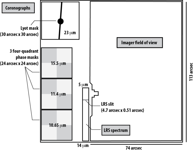
The Mid-Infrared Instrument (MIRI) on the James Webb Space Telescope (JWST) will provide direct imaging and medium resolution spectroscopy (R~ 1500 to 3500) over the wavelength range 5 - 28.5 micron, coronagraphic imaging at 10.65, 11.4, 15.5 and 23 micron, and low-resolution spectroscopy (R~100) over the wavelength range 5 - 12 micron (with some response to 14 micron).
This figure shows the layout of the direct imaging, coronagraphic imaging and low-resolution spectroscopy. Medium resolution spectroscopy is performed with two other detectors (not represented here).
MIRI uses two types of coronagraphic techniques: Lyot coronagraphy and four-quadrant phase mask coronagraphy.
The coronagraphic masks are located along one edge of the focal plane aperture. They include three four-quadrant phase masks and a focal plane mask for a Lyot coronagraph.
In reality, the four-quadrant phase masks are rotated slightly (by approximately 5 degrees) around their centre positions compared to what is shown in this figure. (This can be clearly seen in this flat field image taken during ISIM CV-3 tests.)
The coronagraphic masks each have a square field of view of about 24" × 24" for the phase masks and about 30" × 30" for the Lyot coronagraph. They are optimized for particular wavelengths (10.65, 11.4, 15.5 and 23 μm) with each coronagraph uniquely associated with a specific filter.
Also shown in this schematic figure are the locations of the 4.7" × 0.51" slit for the low-resolution spectroscopy, and the 113" × 74" field for direct imaging.